J. Biosci. Public Health. 2026; 2(4)

First and Corresponding Author
https://orcid.org/0000-0002-5334-9850
Affiliations:
Department of Medical Laboratory Technology, Poltekkes Kemenkes Jambi, Indonesia
Coauthor
sakdiahsiti@poltekkesjambi.ac.id
https://orcid.org/0009-0002-9815-0308
Affiliations:
Department of Medical Laboratory Technology, Poltekkes Kemenkes Jambi, Indonesia
Background: Diabetes mellitus (DM) is characterized by persistent hyperglycemia and biological processes involving inflammation. Lambak Pipih fish oil, a local resource from Jambi, contains vitamin D3 and omega-3 fatty acids and may have metabolic potential. Objective: This study aimed to descriptively evaluate changes in blood glucose and interleukin-17 (IL-17) levels following administration of Lambak Pipih fish oil in an alloxan-induced DM mouse model. Methods: Thirty male Mus musculus mice were allocated to five groups (n = 6): normal control (K1), diabetic control (K2), and three treatment groups receiving Lambak Pipih fish oil at 100, 200, or 300 mg/kg body weight (K3–K5) for 14 days. Diabetes was induced with alloxan monohydrate (150 mg/kg). Blood glucose was measured on days 0, 7, and 14; IL-17 was assessed at baseline and day 14. Only group-level means were available; results are therefore reported descriptively. Results: By day 14, blood glucose increased from 288.7 to 315.2 mg/dL in the diabetic control group, whereas it decreased from 290.3 to 241.6 mg/dL (16.77%) in K3, from 293.5 to 215.8 mg/dL (26.46%) in K4, and from 289.4 to 202.6 mg/dL (30.00%) in K5. IL-17 increased by 3.23% in the diabetic control group, whereas reductions of 14.16%, 25.82%, and 37.24% were observed in K3, K4, and K5, respectively. The largest descriptive reductions in both outcomes were observed at 300 mg/kg BW. Conclusion: Lambak Pipih fish oil was associated with descriptive reductions in blood glucose and IL-17 levels in the diabetic mouse model, with larger changes observed at higher doses. These preliminary trends require confirmation with individual-level data, chemical characterization of the oil, and appropriate inferential statistics before any translational conclusions can be drawn.
Diabetes Mellitus (DM) is a chronic metabolic disease characterized by hyperglycemia resulting from impaired insulin secretion, insulin resistance, or a combination of both [1, 2]. The International Diabetes Federation estimates about 588.7 million adults aged 20–79 years were living with diabetes in 2024, and is projected to increase to 852.5 million by 2050 [3]. Almost 81% of diabetes patients are from low- and middle-income countries [4]. In Indonesia, the IDF estimates that approximately 20.4 million adults were living with diabetes in 2024, with an age-standardized prevalence of 11.3%, placing Indonesia among the countries with the highest number of people with diabetes worldwide [5]. In Jambi Province, the prevalence based on physician diagnosis reached 2.19%, which was higher than the provincial figure [6].
Inflammation is an important mechanism underlying the development and progression of DM because increased levels of pro-inflammatory mediators, such as tumor necrosis factor-alpha (TNF-α), interleukin-1β (IL-1β), and interleukin-6 (IL-6), may exacerbate insulin resistance, tissue damage, and the risk of microvascular complications [7]. Therefore, DM management should not be limited to reducing blood glucose levels but should also consider the inflammatory mechanisms underlying disease progression that may be attenuated through natural-based approaches, particularly the use of nutrient sources with immunomodulatory and anti-inflammatory activities [8].
Two nutrients with complementary immunomodulatory and metabolic actions have attracted substantial attention. Vitamin D, acting through the vitamin D receptor (VDR), suppresses Th17 polarization, reduces IL-17 production, enhances regulatory T-cell function and dampens NF-κB signalling [9]. Meanwhile, omega-3 fatty acids, particularly eicosatetraenoic acid (EPA) and docosahexaenoic acid (DHA), may influence eicosanoid metabolism, reduce the production of pro-inflammatory mediators, modulate leukocyte migration, and contribute to improved insulin sensitivity [10]. The combination of vitamin D and omega-3 activities theoretically provides a strong biological rationale for developing interventions capable of targeting two important components of DM simultaneously, namely impaired glucose regulation and inflammation [11].
Therefore, Lambak Pipih fish oil is of particular interest because it is a local resource that naturally contains both of these components [11]. Previous research on Lambak Pipih fish oil from Jambi has reported substantial vitamin D₃ content and a lipid profile containing omega-3 fatty acids, providing a rationale for further experimental evaluation of this local resource [12]. Some studies have also reported potential effects on metabolic and inflammatory biomarkers, but these findings depend on the source, composition, dose, and experimental context [13, 14]. The relationship between administration of the oil and biological responses under hyperglycemic conditions has not been adequately elucidated [11]. Although evidence regarding vitamin D and omega-3 fatty acids as individual components has continued to develop [15,16], these findings cannot automatically be generalized to Lambak Pipih fish oil because the effects of natural products may be influenced by composition, fish source, extraction process, concentration of active components, and administered dose [13,17].
Moreover, Previous studies have characterized its vitamin D₃ content and lipid profile [18], yet no controlled evaluation of its impact on blood glucose or IL-17 in a diabetic model has been reported. Effects of natural fish oils are known to vary with species, extraction method, dose and batch composition [19, 20]. therefore, findings from purified EPA/DHA or other fish oils cannot be automatically induced. Although the individual effects of vitamin D and n-3 PUFAs are increasingly well documented [21, 22]. experimental evidence for Lambak Pipih fish oil itself remains scarce. In addition, evidence regarding the biological effects of Lambak Pipih fish oil in DM remains very limited.
The present study integrates the potential of two major bioactive components, namely vitamin D and omega-3 fatty acids, within a single local natural resource, Lambak Pipih fish oil. This approach is directed not only toward controlling hyperglycemia but also toward examining changes in IL-17, an inflammatory cytokine relevant to the biological response associated with diabetes. Moreover, it evaluates the effects of orally administered Lambak Pipih fish oil at three doses (100, 200 and 300 mg/kg body weight) for 14 days on blood glucose and circulating IL-17 in an alloxan-induced diabetic mouse model. The study hypothesis is that administration of Lambak Pipih fish oil will reduce blood glucose levels and attenuate inflammatory responses in diabetic model mice more effectively than the negative control group, with responses varying according to the administered dose.
2.1. Study design
This was a controlled, repeated-measures, in-vivo laboratory experiment conducted in male Mus musculus mice. Animals were allocated to five groups (n = 6 per group): normal control (K1), diabetic control (K2), and three treatment groups that received Lambak Pipih fish oil at 100 mg/kg (K3), 200 mg/kg (K4) or 300 mg/kg body weight (K5). Blood glucose was measured on days 0, 7 and 14; circulating IL-17 was measured at baseline and at the end of the 14-day intervention.
2.2. Animals and housing
Thirty male white mice (Mus musculus), 2–3 months old and weighing 25–30 g, were obtained from a commercial supplier and housed at the Animal Laboratory, Faculty of Medicine and Health Sciences, Universitas Jambi. Animals were maintained under controlled conditions (25 ± 2 °C, 12 h light/dark cycle) with free access to standard chow and water. After a 7-day acclimatization period they were fasted for 12 h before diabetes induction.
2.3. Induction of diabetes
Diabetes was induced by a single intraperitoneal injection of alloxan monohydrate (150 mg/kg body weight) dissolved in sterile 0.9 % saline. Seventy-two hours later, blood was collected from the tail vein and glucose concentration was determined by an enzymatic method. Only mice with fasting blood glucose ≥ 200 mg/dL were retained as diabetic models and entered the treatment phase. Group K1 received vehicle only and served as the non-diabetic control.
2.4. Preparation and administration of Lambak Pipih fish oil
Whole fresh Lambak Pipih fish (30 kg) sourced from Jambi Province were processed by sequential heating (60 °C then 95 °C), mechanical pressing and centrifugation (5 000 rpm, 25 °C, 10 min). The resulting oil was stored in amber glass bottles at 4 °C until use. Fish oil was administered once daily by oral gavage for 14 consecutive days at the doses stated above. Control groups (K1 and K2) received an equivalent volume of distilled water. Batch-specific quantification of vitamin D₃, EPA, DHA or total omega-3 content was not performed on the oil used in this experiment; previously published compositional data for Lambak Pipih oil therefore cannot be assumed to apply to the present batch.
2.5. Outcome measures
Blood glucose. Tail-vein blood was collected on days 0 (baseline), 7 and 14 after a 12-h fast and analyzed by a standardized enzymatic method (Clinical Chemistry Laboratory, Poltekkes Kemenkes Jambi). Results are expressed in mg/dL.
Interleukin-17. Circulating IL-17 was measured at baseline and on day 14. Because the original laboratory records did not contain the kit manufacturer, catalogue number, detection limit or intra-/inter-assay coefficients of variation, these analytical performance parameters cannot be reported.
2.6. Sample size and allocation
Sample size (n = 6 per group) was determined by the pre-defined experimental protocol and resource constraints; no formal a-priori power calculation was performed. Allocation of animals to the five groups followed the protocol sequence; formal randomization and allocation concealment were not documented. Investigators performing outcome measurements were not blinded to group assignment.
2.9. Data analysis
The available dataset contained group-level means without individual-level observations or measures of variability. Therefore, the analyses reported in this manuscript were limited to descriptive statistics: group means at each measurement time point and percentage change from baseline to day 14. No p-values, confidence intervals, effect sizes, or inferential comparisons were calculated from the available data. Accordingly, the findings are presented as descriptive trends rather than statistically demonstrated treatment effects.
3.1. Blood glucose levels
Based on Table 1, blood glucose levels in the normal control group remained relatively stable, decreasing from 103.2 mg/dL on day 0 to 101.4 mg/dL on day 14. In the diabetic control group, blood glucose increased from 288.7 mg/dL to 315.2 mg/dL. In contrast, the treatment groups showed descriptive reductions to 241.6 mg/dL at 100 mg/kg BW, 215.8 mg/dL at 200 mg/kg BW, and 202.6 mg/dL at 300 mg/kg BW. The corresponding baseline-to-day-14 reductions were 16.77%, 26.46%, and 30.00%, respectively. Because variability and individual-level observations were not available, these differences cannot be interpreted as statistically significant treatment effects.
Table 1. Mean blood glucose levels of mice by treatment group.
| Group | Day 0 (mg/dL) | Day 7 (mg/dL) | Day 14 (mg/dL) |
| K1 | 103.2 | 106.1 | 101.4 |
| K2 | 288.7 | 301.6 | 315.2 |
| K3 | 290.3 | 263.8 | 241.6 |
| K4 | 293.5 | 247.1 | 215.8 |
| K5 | 289.4 | 229.7 | 202.6 |
Note: K1 = normal control; K2 = diabetic control; K3 = fish oil 100 mg/kg BW; K4 = fish oil 200 mg/kg BW; K5 = fish oil 300 mg/kg BW.
As presented in Figure1, blood glucose levels decreased from day 0 to day 14 by 16.77% in K3, 26.46% in K4, and 30.00% in K5. The magnitude of reduction was numerically larger at higher doses; however, this pattern should be regarded as a descriptive dose-related trend rather than a statistically confirmed dose-response relationship.
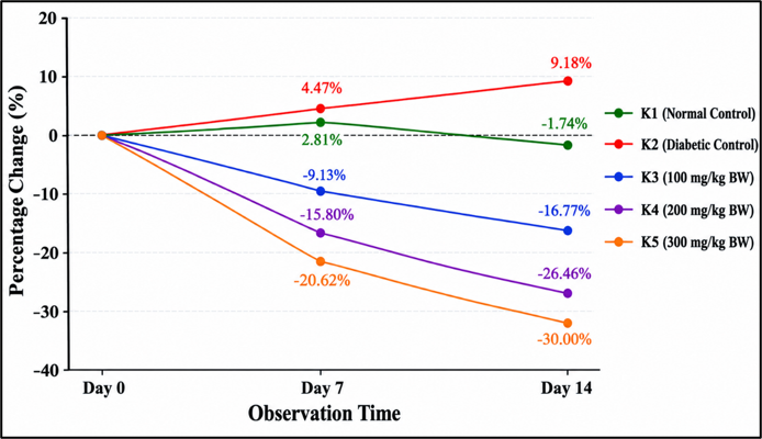
Figure 1. Percentage change in the measured parameter across the 14-day observation period in the normal control (K1), diabetic control (K2), and treatment groups receiving 100, 200, and 300 mg/kg body weight (BW) (K3–K5).
IL-17 levels before and after administration of Lambak Pipih fish oil are presented in Figure 2. The figure should be based on the verified IL-17 dataset and should not contain blood-glucose values. Because the individual-level IL-17 data and variability measures were not available in the manuscript dataset, the current text reports only the group-level changes that were available. To facilitate visualization of changes in IL-17 levels across the treatment groups, the IL-17 levels before and after administration of Lambak Pipih fish oil are presented in Figure 2. The graph illustrates the pattern of changes in IL-17 levels in each group, facilitating comparison of inflammatory responses between the control groups and the intervention groups.
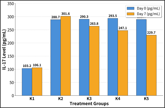
Figure 2. IL-17 Levels before and after administration of Lambak Pipih fish oil.
3.2. IL-17 levels
Based on the available IL-17 results, the K1 normal control group showed a small increase in IL-17 levels from 117 pg/mL on day 0 to 121 pg/mL on day 14, corresponding to a 3.42% increase (Figure 3). The K2 diabetic control group also showed an increase, from 238.6 pg/mL to 246.3 pg/mL, corresponding to a 3.23% increase. In contrast, the fish-oil treatment groups showed descriptive reductions in IL-17 levels of 14.16%, 25.82%, and 37.24% in K3, K4, and K5, respectively (Figure 3).
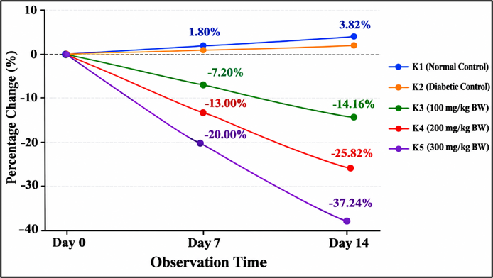
Figure 3. Percentage change in the measured parameter across the 14-day observation period in the normal control (K1), diabetic control (K2), and treatment groups receiving 100, 200, and 300 mg/kg BW (K3–K5), showing dose-dependent reductions in the treatment groups.
The higher IL-17 level observed in K2 than in K1, together with the descriptive reductions observed in the fish-oil treatment groups, indicates a pattern of change in IL-17 levels during the experimental period. The magnitude of the reported reduction increased across the 100, 200, and 300 mg/kg BW doses. However, because the available dataset contains group-level results without individual-level observations or measures of variability, no statistical significance can be established. Therefore, these findings should be interpreted as descriptive trends rather than statistically confirmed treatment effects. The present findings should also be interpreted specifically in relation to IL-17. Although IL-17 is an inflammatory cytokine and may be involved in immune dysregulation associated with diabetes, it represents only one component of the broader inflammatory network. Therefore, the observed reduction in IL-17 should not be interpreted as evidence of a generalized reduction in inflammation or as definitive evidence of an anti-inflammatory effect of Lambak Pipih fish oil.
This study was designed to describe changes in blood glucose and IL-17 following administration of Lambak Pipih fish oil in an alloxan-induced Diabetes Mellitus model. The diabetic control group showed an increase in blood glucose from 288.7 mg/dL to 315.2 mg/dL after 14 days, whereas the fish-oil groups showed descriptive reductions of 16.77%, 26.46%, and 30.00% at doses of 100, 200, and 300 mg/kg BW, respectively. IL-17 also showed reported reductions of 14.16%, 25.82%, and 37.24% at the corresponding doses. These patterns are numerically consistent with greater changes at higher doses. However, because individual-level observations and variability measures were not available, no p-values, confidence intervals, or effect sizes could be calculated. The findings therefore represent descriptive trends requiring statistical confirmation rather than evidence of statistically established effectiveness.
The observed reductions in blood glucose and IL-17 after 14 days of Lambak Pipih fish oil administration, relative to the progressive rise in the diabetic control group, are consistent with a favorable metabolic and inflammatory response in this alloxan-induced model. Because analyses were limited to group means, these patterns remain exploratory trends and cannot be interpreted as statistically confirmed treatment effects.
The direction of change aligns with meta-analytic evidence that n-3 polyunsaturated fatty acid (PUFA) supplementation can favorably influence glycemic and inflammatory markers in type 2 diabetes, although effect sizes and biomarker specificity vary across trials [23–25]. Heterogeneity remains substantial and is influenced by dose, duration, EPA:DHA ratio, baseline inflammatory status and the specific biomarker assessed [26]. The present experiment differs from human trials in model, intervention matrix (whole fish oil rather than purified EPA/DHA), dose, duration and exclusive focus on IL-17; therefore, directional consistency should not be interpreted as quantitative equivalence.
Persistent hyperglycemia promotes oxidative stress and NF-κB activation, increasing transcription of pro-inflammatory cytokines [7]. EPA and DHA remodel membrane phospholipids, alter eicosanoid synthesis, attenuate NF-κB signaling and generate specialized pro-resolving mediators [8]. Vitamin D, via the vitamin D receptor, suppresses Th17 polarization, reduces IL-17 production, enhances Treg function and dampens NF-κB-driven inflammation [9,15]. The concurrent decline in glucose and IL-17 is biologically plausible under these pathways. However, the experimental oil was not assayed for EPA, DHA or vitamin D₃; mechanistic attribution therefore remains hypothetical. IL-17 represents only one node in a complex cytokine network and cannot be extrapolated to a global anti-inflammatory effect [27].
The larger numerical changes at higher doses are consistent with a descriptive dose-related pattern but do not constitute formal evidence of a dose–response relationship. Prior rodent studies using fish oils in alloxan-induced diabetes have reported graded glycemic improvements, supporting biological plausibility [28, 29]. Lambak Pipih fish oil is a locally available resource in Jambi that merits further characterization. The present findings provide only preliminary experimental information. Translation to clinical use would require batch-specific chemical standardization, safety evaluation, adequately powered individual-level studies, additional metabolic and inflammatory endpoints, and well-designed human trials.
Key limitations include the exclusive use of group-level means (precluding inferential statistics), lack of formal power calculation and randomization documentation, absence of batch-specific chemical characterization, incomplete IL-17 assay details, and the short 14-day duration in an alloxan model. These factors classify the results as exploratory descriptive trends requiring confirmation in more rigorous designs.
Future studies should employ justified sample sizes, repeated-measures analyses, full chemical characterization of the oil, complete assay reporting, and broader biomarker panels before any translational claims are considered.
Oral administration of Lambak Pipih fish oil for 14 days was associated with descriptive, dose-related reductions in mean blood glucose and IL-17 levels in an alloxan-induced diabetic mouse model, with the largest numerical changes observed at 300 mg/kg BW. These preliminary trends are directionally consistent with existing evidence on omega-3 fatty acids and vitamin D but do not establish statistically significant treatment effects or a confirmed dose–response relationship. Future studies should employ individual-level data, justified sample sizes, batch-specific chemical characterization, complete assay reporting, additional metabolic and inflammatory endpoints, and appropriate safety assessment before any translational or clinical application is considered.
The authors would like to express their gratitude to Poltekkes Kemenkes Jambi, for providing facility and technical support throughout the study. Appreciation is also extended to all members of the research team and laboratory technicians who assisted with experimental animal maintenance, intervention procedures, sample collection, laboratory examinations, and research data management. The authors also extend their sincere thanks to all parties who provided support and contributed to the successful completion of this study.
This research received no specific grant from any funding agency.
The authors declare that they have no conflicts of interest.
The animal study was conducted in accordance with animal welfare principles and applicable ethical standards for experimental research involving laboratory animals. The study used the minimum number of animals included in the experimental protocol, monitored animal condition throughout the intervention, and applied appropriate handling during intervention administration and biological sample collection. Ethical approval was obtained from the Health Research Ethics Committee of Poltekkes Kemenkes Jambi, Indonesia, under Ethical Approval No. LB.02.06/2/478/2026.
The data that support the findings of this study are available in the article. Further details may be obtained from the corresponding author upon request.
AI tools were used for language editing, grammar correction, and improving the readability and structure of this manuscript in accordance with the journal's policy. No AI tools were used to generate data, fabricate references, or create figures/tables.

This work is licensed under a
Creative Commons Attribution 4.0 International License
.
You are free to share and adapt this material for any purpose, even commercially, as long as you give appropriate credit.
 access